Diagnostic imaging · dogs and cats
Magnetic resonance imaging
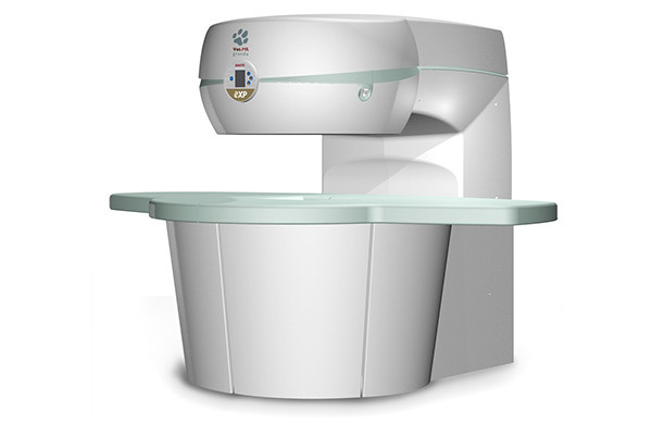
Magnetic resonance imaging (MRI) is a non-invasive examination used to assess internal organs and tissues. In veterinary medicine, it is most commonly used to diagnose diseases of the central nervous system — the brain and spinal cord.
MRI is also frequently used to diagnose disorders of the skeleton, muscles, tendons and joints. It is one of the most precise imaging examinations in both human and veterinary medicine.
Indications
MRI is most commonly performed as part of planned and preventive diagnostics for diseases of the central nervous system and tumours.
Scope of the examination
The examination can visualise a range of organs and structures:
- spine: investigation of spinal pain (all spinal regions), assessment of muscles and ligaments, and evaluation of vertebral disorders (inflammation, tumours, metastases and degenerative changes)
- head: imaging of disorders affecting the brain, ears, lymph nodes and salivary glands
- limbs and joints: assessment of the knee, shoulder, elbow and hip joints, ligaments, and inflammatory, degenerative and neoplastic changes in bone structures.
Preparing for the scan
The patient must fast for at least 8 hours.
Remove access to water 3 hours before anaesthesia.
Before anaesthesia, basic blood tests and a cardiac assessment are required. Patients with diagnosed heart disease or other chronic conditions (diabetes, kidney or liver disease) should have an anaesthesia consultation before the procedure.
Auxilium · Warsaw
Book an appointment
Choose an appointment online or contact our reception team.